43+ Vascular Bundle Monocot Root Cross Section Pictures. Barnes does attempt to bust out some tutorials but does not claim to know. Diagram illustrating the tissue layers and their organization within monocot and dicot roots.
Dicot Stem Vs Monocot Stem What Is The Difference Diffzi from diffzi.com
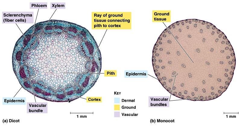
In some monocot roots the hypodermis (exodermis) is also heavily sclerenchymized. Note the ringed array of vascular bundles in this zea (monocot) root cross section. Monocots and dicots differ from each other in four structures:
Monocot roots are fibrous and networked 2.
The xylem system consists of tracheids. The vascular tissue is in the very center of the note the sclerenchymized endodermis and epidermis. In monocot roots, the vascular bundles are arranged in a circular pattern. The vascular system found in dicots is somewhat more complex than that found in monocots.